


|

|


Lyle Gordon, Derk Joester
______________________________________________________________
Overview
Biological organisms have a remarkable ability to control the structure and properties of inorganic minerals across a wide range of length scales from nano- to macro-scale[1]. In many cases, an organic matrix comprised of a variety of macromolecules interacting with the forming mineral is responsible for the control over the morphology and the polymorph of the mineral. For example, in the tooth of the chiton the mineralization occurs within a preformed organic matrix. This organic matrix, composed of the polysaccharide chitin and several proteins, is responsible for controlling the mineral deposition[2][3]. As the tooth matures, the organic matrix is incorporated into the forming mineral. The outstanding fracture toughness and wear resistance of the tooth results from the organic-inorganic interfaces over multiple levels of hierarchy (i.e. intra- and inter-crystalline) that deflect and arrest cracks[4]
An in-depth understanding of nucleation, growth, and the advanced material properties resulting from interfaces at several length scales depends on the ability to characterize the interactions across these organic-inorganic interfaces. Atom probe tomography (APT) is uniquely capable of providing structural insight at the atomic scale by directly mapping the location and chemical identity of the atoms within a small sample of material, usually a metal or semi-conductor[5].
An atom probe is a point projection microscope where, in the presence of a very high electric field, individual atoms or molecules sequentially field-evaporate from a sharp tip and are projected onto a 2D position sensitive detector (Scheme) The mass-to-charge state ratio (m/z) and thus the chemical identity of each ion are determined by time-of-flight (TOF) mass-spectrometry using timed picosecond laser pulses to trigger evaporation events. In combination with the sequence of evaporation events and their location on the detector, this allows reconstructing the 3D structure of the sample, atom by atom. APT analyzes volumes on the order of 105 nm3 with sub-nanometer resolution.
Recent advances in atom probe technology, including improvements in laser pulsing, have enabled the atom probe to study bulk materials with high resistivity. Examples include numerous metal and semiconductor oxides [6-9] and polymers [10]. We use a pulsed laser atom probe was used to characterize the buried organic-inorganic interfaces within biological minerals. We demonstrated the applicability of the technique using the tooth cap of the chiton (Figure 1A-B) a magnetite-organic composite, containing numerous nanoscale organic fibers (Figure 1C),. 3D Atom probe reconstructions of the chiton tooth (Figure 1D) reveal the organic fibers and enable sub-nanometer resolution chemical mapping of the inorganic-organic interface. We are currently working to extend the approach to other biomineral systems where organic/inorganic interfaces play an important role.
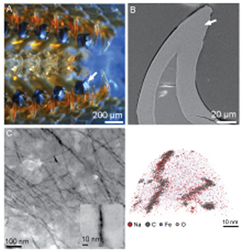
Figure 1
References
[1] Lowenstam, H. & Weiner, S. On biomineralization, (Oxford University Press, USA, 1989).
[2] Evans, L.A., Macey, D.J. & Webb, J. Characterization and Structural Organization of the Organic Matrix of the Radula Teeth of the Chiton Acanthopleura hirtosa. Philosophical Transactions of the Royal Society B: Biological Sciences 329, 87-96 (1990).
[3] Evans, L., Macey, D. & Webb, J. Distribution and composition of matrix protein in the radula teeth of the chitonAcanthopleura hirtosa. Marine Biology 109, 281-286 (1991).
[4] van der Wal, P., Giesen, H.J. & Videler, J.J. Radular teeth as models for the improvement of industrial cutting devices. Materials Science and Engineering: C 7, 129-142 (2000).
[5] Miller, M.K. Atom Probe Tomography: Analysis at the Atomic Level, (Plenum Pub Corp, 2000).
[6] Oberdorfer, C., Stender, P., Reinke, C. & Schmitz, G. Laser-Assisted Atom Probe Tomography of Oxide Materials. Microscopy and Microanalysis 13, 342-346 (2007).
[7] Larson, D.J., et al. Analysis of Bulk Dielectrics with Atom Probe Tomography. Microscopy and Microanalysis 14, 1254-1255 (2008).
[8] Chen, Y., Ohkubo, T., Kodzuka, M., Morita, K. & Hono, K. Laser-assisted atom probe analysis of zirconia/spinel nanocomposite ceramics. Scripta Materialia 61, 693-696 (2009).
[9] Kuhlman, K.R., Kelly, T.F. & Miller, M.K. Atomic-Scale Analysis of Metamorphic Magnetite using Field Ion Microscopy and Atom Probe Tomography. Microscopy and Microanalysis 10, 512-513 (2004).
[10] Evans, J.E., et al. 3-D Atom Probe Tomography of Resin Embedded Samples? Microscopy and Microanalysis 15, 274 (2009).

Home | Contact Us | Sites A-Z